The Electric Consensual Response of the Non-stimulated Eye in Normal Subjects and Patients with Optic Atrophy
P. Steindler, P. Cardin and S. Perrone
Eye Clinic of the University of Padova, Via Giustiniani, I-35100, Padova, Italy
Abstract
The electric consensual response wlien the collateral eye underwent electroretinographic stimulation was studied. Results obtained from normal subjects and 17 patients with optic atrophy are discussed.
Introduction
Many studies have been conducted in humans and in other animals to verify whether the stimulation with light of one retina could in some way influence the electrical activity of the controlateral retina. Motokawa and Mita (1942), Karpe (1945), Monnier (1946), and Dodt (1956) observed a positive or negative electrical potential in the controlateral retina with a latency of 150-300 ms.
Uchermann (1955) and Bagolini (1959) could not demonstrate a consensual electroretinographic response in man, whereas Wirth (1951) noted that the bwave of the ERG was significantly smaller with binocular stimulation than with monocular stimulation. Muller-Limmroth (1954) observed a positive electrical potential in the non-illuminated eye of the guinea pig with a delay of 50 ms. This potential did not appear after section of the optic nerve or destruction of the lateral geniculate body on the side of the non-stimulated eye. The author, therefore, hypothesized the presence of crossed pathways in the chiasma connecting the two retinae.
Auerbach et al. (1961) and Hosten et al. (1961) observed, in humans and animals, synchronized potentials of opposite polarity to the b-wave of the non stimulated eye. These authors, however, later hypothesized that such potentials could represent simple diffusion of electric current from the stimulated eye through the surrounding tissues.
Ponte and Monaco (1963), in a series of experiments in the rabbit, reached the same conclusion. In spite of the fact that there have been a series of anatomical (Cajal 1894; Polyak 1941; Wolter 1960; Cragg 1962; Pfister 1963) and physiological demonstrations (Granit 1955; Dodt 1956; Hunt and Jakobson 1972; Dowlingetal 1976) of the presence of centrifugal nervous path ways in the optic nerves, this hypothesis is far from being clear.
Because of the controversy over such an important physiological problem, it was felt worthwhile to repeat this type of study in man. In particular, the existence of consensual potentials in normal subjects and in subjects with both unilateral and bilateral optic atrophy was verified. In the latter case, the presence of potentials in the non-stimulated eye was not possible without a simple diffusion through the tissues.
Subjects and Methods
This study was conducted with 20 normal subjects and in 17 subjects with optic atrophy of various types; eight cases were unilateral and ten bilateral. Unilateral stimulation was first applied to the right eye and then to the left. Such stimulation was carried out in scotopic adaptation in eyes in which mydriasis was obtained with Visumidriatic (1%) eye drops. A Vescovini model 481 stimulator using a Xenon lamp (3 J) with white light and a frequency of one flash per second was employed.
Unilateral stimulation was effected using a type of underwater goggle which fitted perfectly to the anatomy under which corneal electrodes (Henkes type) and cutaneous electrodes (Fig. 1) were placed. The corneal electrode was connected with the positive input of the pre-amplifier.
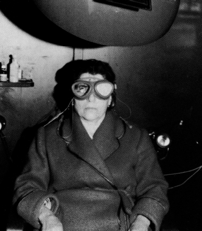
Fig. 1. Position of the patient during examination inside Faraday's cage
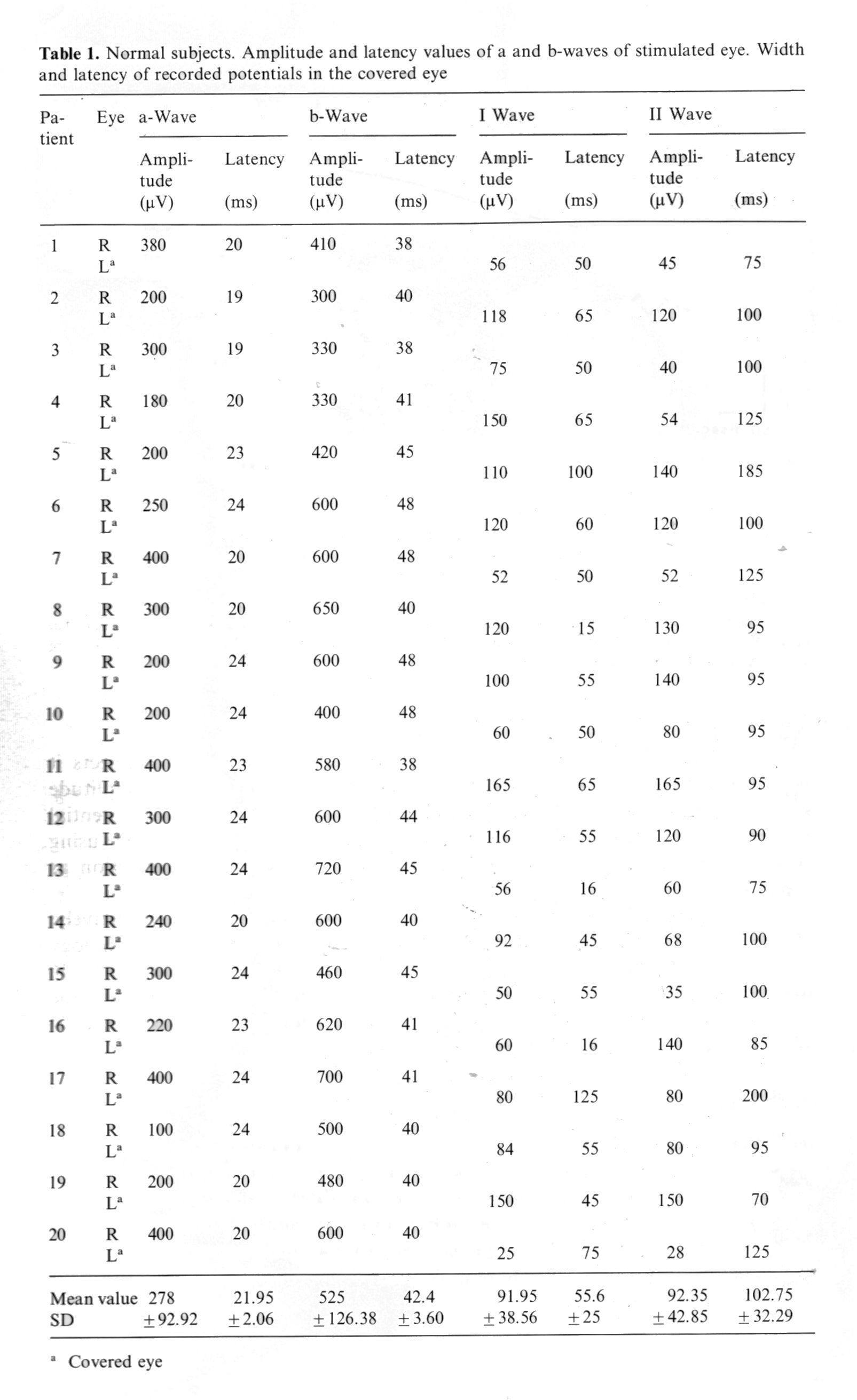
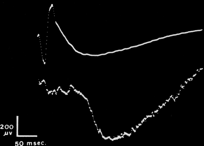
Fig. 2. Above: ERG of the stimulated eye. Below: Electric consensual response of the non-stimulated eye
The potentials thus obtained were amplified by a Vescovini pre-amplifier with a passing hand from 0.3-500 Hz/s fed into a Packard Averager (Model 3240) so that each tracing derived from the average of 16 stimulations.
Visual-evoked potentials with a unipolar derivation using an occipital electrode placed on the median line 3 cm above the inion were simultaneously recorded. The occipital electrode was connected with the negative input of the pre-amplifier, the indifferent electrode on the ear lobe being connected to the positive input.
Results
The data obtained are summarized in Table 1. In all the normal subjects it was possible to show, in the non-stimulated eye, a potential with an amplitude of 25-200 µV after stimulation of the controlateral eye (Fig. 2). This potential was composed of two positive deflections with amplitudes calculated by using the base line passing through the apex of the negative inferior deflection as a reference.
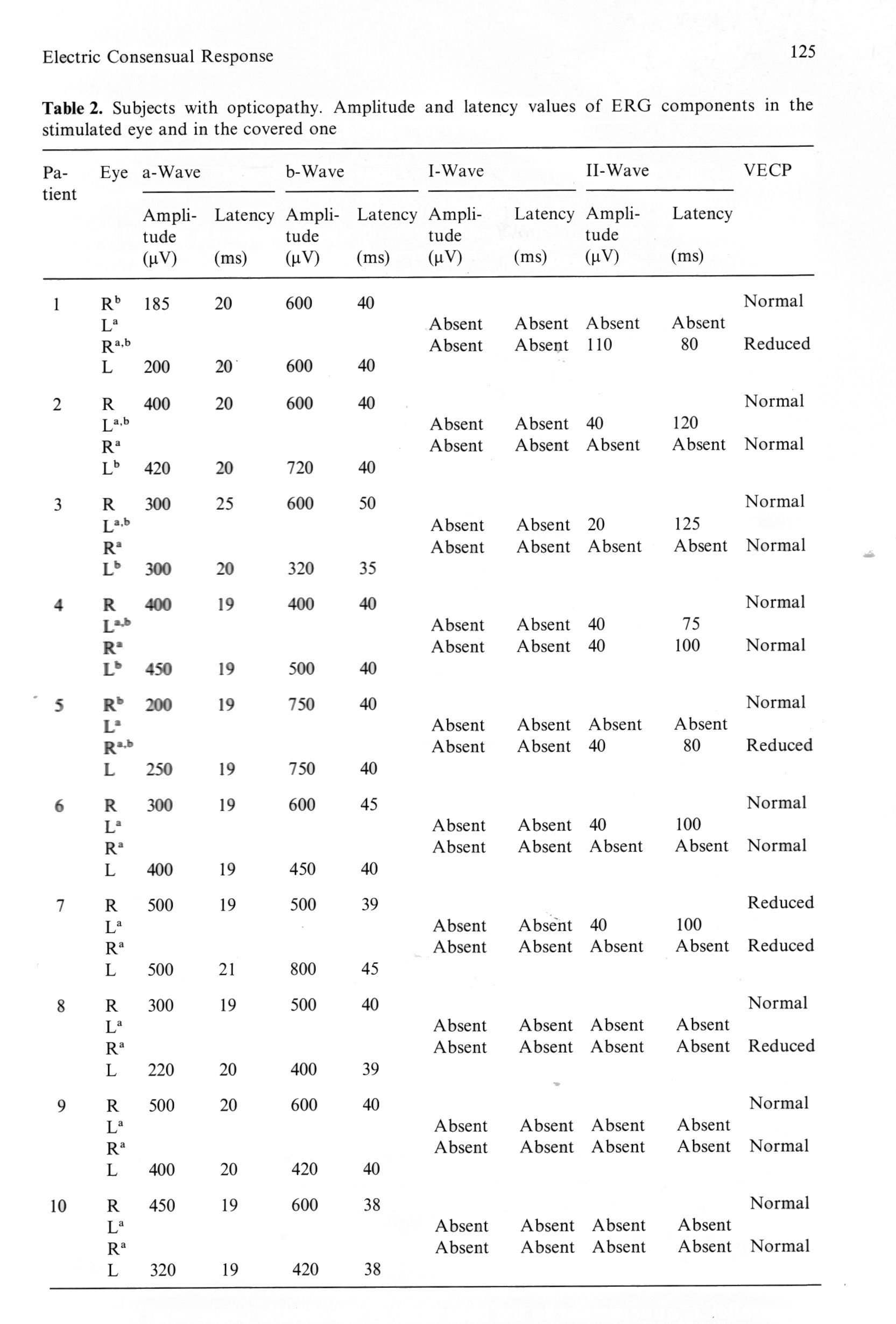
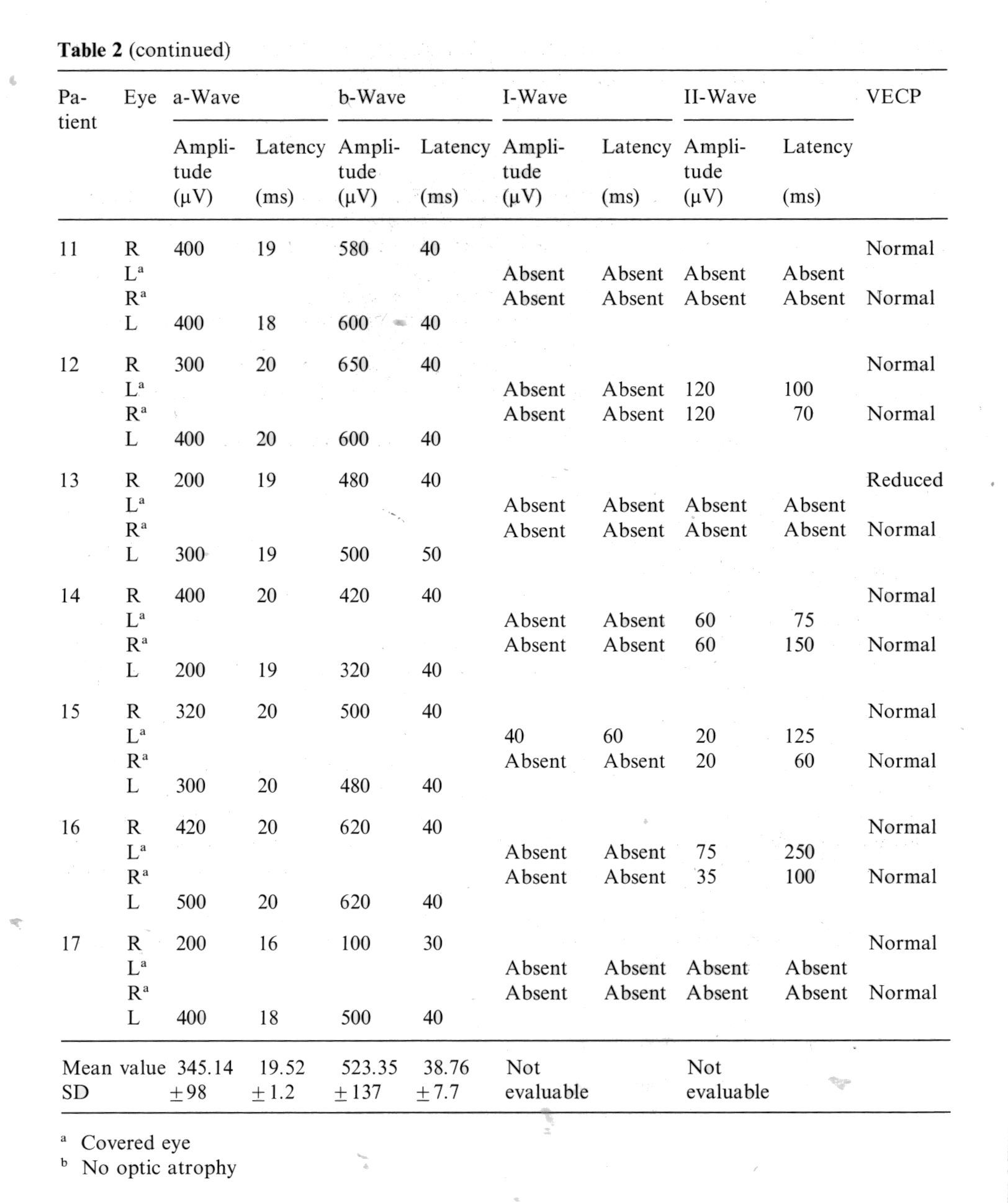
The average amplitude of waves I and II was 91.5 and 92.5 µV respectively. The results of the stimulation carried out in subjects with optic nerve pathology are shown in Table 2. In the more severe bilateral cases (patients 8, 9, 10 and 11), the consensual response appears to be completely abolished with the VER. In the unilateral, cases a net reduction or complete disappearance of the consensual potential was observed.
Discussion
The results obtained in normal subjects clearly demonstrate the presence of a recordable potential in the eye in relation to light stimulation of the consensual eye. With this type of experiment, it is not possible to demonstrate whether these potentials are due to biretinal interactions transmitted through the optic nerve or to a transmission through pathways other than nervous ones i.e. by simple diffusion through surrounding tissues.
The matter is clarified if the results obtained in subjects with optic atrophy are examined. In such cases, consensual potentials are drastically reduced in unilateral optic atrophy, or absent in several cases of bilateral optic atrophy. This would favour the hypothesis that the diffusion from one eye to the other occurs via structures contained in the optic nerve.
This assumption can be confirmed by the behaviour of VECP which, though not parallel, is quite similar to the behaviour of the electric consensual response. In fact, a reduction or absence of the consensual potential corresponds in most cases to a reduction, or to an absence, of VECP.
References
-
Auerbach E, Beller AJ, Henkes HE, Goldhaber G (1961) Electric potentials of retina and cortex of cats evoked by monocular and binocular photic stimulation. Vision Res 1: 166 - 182
-
Bagolini B (1959) L'elettroretinogramma durante stimolazioni monoculari e binoculari. Boll Ocul 38: 605 -615
-
Cajal SR (1894) Die Retina der Wirbelthiere. Bergmann, Wiesbaden
-
Cragg BG (1962) Centrifugal fibers to the retina and olfactory bulb, and composition of the supraoptic commissures in the rabbit. Exp Neurol 5: 406 - 427
-
Dodt E (1956) Centrifugal impulses in rabbit's retina. J Neurophysiol 19: 301 - 307
-
Dowling JF, Ehinger B, Hedde WL (1976) The interplexiform cell: A new type of retinal neuron Invest Ophthal 15: 916 - 926
-
Granit R (1955) Centrifugal and antidromic effects on ganglion cells of retina. J Neurophysiol 18: 388 - 411
-
Hosten GPM. Wildeboer-Venema FN, Winkelman JE (1961) Studies on centrifugal effects on the retina: a corneonegative potential in a non-illuminated eye. Arch Phys et Bioch 69: 431 - 442
-
Hunt RK. Jakobson M (1972) Development and stability of positional information in xenopus retinal ganglion cells. Proc Nat Ac Sci U.S.A. 69: 4
-
Karpe G (1945) The basis of clinical electroretinography. Acta Ophthal Suppl 24
-
Monnier M (I946) Les manifestations electriques consensuelles de l'activite retinienne chez l'omme. (Electroretinographie binoculaire). Experientia 2: 190 - 191
-
MOTOkawa K, Mita T (1942) Uber eine einfachere Untersuchungsmethode und Eigenschaften der ktionsstrome der Netzhaut des Menschen. Tohoku J Exp Med 42: 114 - 133
-
Muller-Limmroth HW (1954) Elektrophysiologische Untersuchungen zum Nachweis einer biretina- len Association. Z Biol 107: 216 - 240
-
Pfister RR, Xolte JR (1963) Centrifugal fibers of the human optic nerve. Neurology 13: 38 - 42
-
Polyak SL (1941) The retina. Chicago Univ Press, Chicago
-
Ponte F, Monaco P (1963) Studio delle modificazioni indotte dalla stimolazione luminosa monocu lare sull'attivita bioelettrica della retina e del nervo ottico controlaterali. Giorn It Oftalm 16: 1 - 14
-
Uchermann A (1955) The electroretinogram on binocular and monocular light stimulation. Acta Ophthal 33: 517 - 522
-
Wirth A (1951) Nota sul meccanismo dei riflessi interretinici. Boll Ocul 30: 499 - 504
-
Wolter JR (1960) Regenerative potentialities of the centrifugal fibers of the human optic nerve. AMA Arch Ophthal 64: 697 - 707

